Dr. Gary Sewell, M.D. B.Sc.
Unicity Eye Clinic
P (204) 953-5560
F (204) 949-1143
Caring For Your Eyes For Over 30 Years
copyright 2022
Dr. Gary Sewell
710 Pembina Highway
Winnipeg, MB
R3M 2M6
P 204 953 5560
F 204 949 1143
KEY POINTS
• A retinal photograph allows a doctor to see and document areas of damage inside the back of the eye
• The retina is the light sensitive tissue lining the inside of the back of the eye
• Damage to the retina from diabetes can cause irreversible vision loss
• The Optomap gives the widest view possible of the back of the eye in one panoramic image allowing the early detection of complications from diabetes that would otherwise be missed
Optomap
The optomap ultra-widefield retinal image is a unique technology that captures more than 80% of your retina in one panoramic image (compared to the 12% of traditional imaging methods).
WHAT HAPPENS DURING AN OPTOMAP TEST?
Having an Optomap is a quick and painless. Nothing touches the eye, there are no air puffs, and it is pain free. It is suitable for people of all ages, including children. Optomap images are immediately available on screen. You can see the inside of your own eye. Images are saved and can be used for comparison at your next eye exam.


Dr. Oz demonstrates the Optomap on his program.
WHY OPTOMAP
The retina is the delicate tissue at the back of the eye which sends a picture to your brain enabling you to see. Diabetes damages the retina causing vision loss. A damaged retina is like a camera with no film.
A retinal photograph allows a doctor to see and document areas of hemorrhage and leakage inside the back of the eye. A standard retinal photograph is very limited in view, showing about 10-12% of the retina. The Optomap ultra-wide digital retinal image captures about 84% of your retina in one panoramic image. This in incredibly important as it allows the early detection of complications from diabetes that would otherwise be missed and it allows an exact record that can be compared against at future visits to say with confidence whether there is improvement or progression.
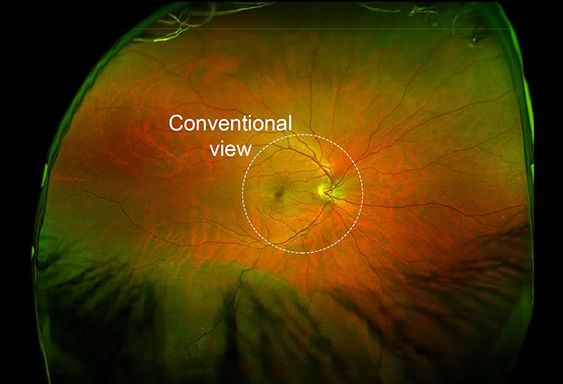
An Optomap image and the area captured by a traditional retinal photograph.
Because the eyeball is a sphere and the Optomap image is so wide, the edges of the
image are actually around the sides, closer to the front of the eyeball.
ARE ALL OPTOMAP TESTS THE SAME?
All Optomaps are not the same. Like any technology the Optos scanner has undergone constant change and refinement and Optos offers a number of qualities of scanner. Only the newest, most advanced machine has a resolution of 14 microns (14 thousands of a millimetre) and it is only this machine that has been proven in the literature to be superior to traditional methods in detecting diabetic retinopathy. As of the time of this writing we are using one of only 3 such machines in Manitoba.

Download a copy of the Optomap brochure.